Hair ties, a keychain, spare change, a Band-Aid…all items that likely reside in your “junk drawer” at this very moment. As animal health professionals know all too well, however, it’s just as likely that these same common household items have ended up inside a pet’s stomach at one time or another. Animals really do eat the darndest things, and that premise is precisely what inspired the infamous “They Ate What?!” radiograph contest, presented annually by Veterinary Practice News.
From a lizard who ingested a rubber Kong toy, to a cat who chowed down on a child’s necklace, to a pup who gobbled up a golf ball (all three animals made full recoveries), there have been some truly remarkable submissions in years past, and we can’t wait to see what this year’s entries will bring. Another reason we’re particularly pumped about the 2023 edition of the contest? We’re sponsoring it! That’s right, Patterson Veterinary and Patterson Teleradiology powered by Vetology AI will be providing the contest prizes, and we couldn’t be more excited about the opportunity to do so.
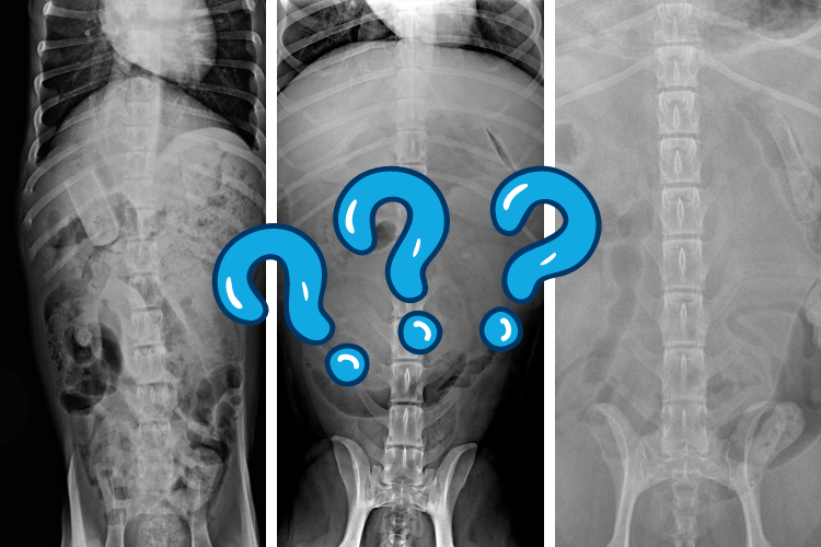
In the spirit of the “They ate What?!” contest, we took to social media and asked you to test your diagnostic skills by sharing how you would read three separate radiographs, each featuring a pet who ate something decidedly other than pet food. Vetology AI, the artificial intelligence software that augments Patterson’s teleradiology services, has been trained on thousands of companion animal cases, resulting in 92% accuracy. However, pets sometimes ingest objects so strange that it even stumps the virtual radiology assistant. In unusual or complicated instances like these, Patterson Teleradiology offers board-certified radiologist overreads for a second opinion. The three cases below represent this type of outlier, and were ultimately reviewed by our radiologists.
Read on for the images, history, summary of findings, conclusions, and recommendations for three intriguing cases. 🤔
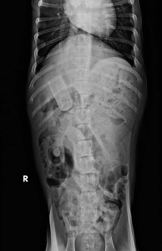
Case A: "Cuddles," a 3-month old female-intact Bernese Mountain Dog
.png "case-a-cuddles1-(1).png")
.png "case-a-cuddles2-(1).png")
History
Ate a 10-inch rawhide around 6 PM, acting normally, has an umbilical hernia.
Radiologist Findings
Abdomen: There is a diffuse reduced peritoneal serosal detail, consistent with age. Within the pyloric region of the stomach, there is a 5.6 x 2.3 cm cylindric smoothly marginated foreign body. This foreign body appears to be handle-or cup- shaped with a rounded closed end, and an open end. The remainder of the stomach is moderately filled with granular food material. The small intestines are variably filled with granular food material, but are within normal limits for shape and size. The transverse and descending colon contains a moderately large amount of feces. The urinary bladder is obscured. The spleen is normal. The kidneys are obscured. The hepatic silhouette is normal.
Conclusions
Gastric foreign body, likely a plastic handle, or cup. A rawhide would be expected to have a more irregular outline and less opaque. The size of the foreign body may predispose for pyloric outflow obstruction, or intestinal obstruction.
Recommendations
Fasting, and endoscopic retrieval, or a laparotomy may be considered. The chances of intestinal obstruction with a conservative approach, are high.
Case B: "Penny," a 10-year old female-intact King Charles Spaniel
.png "case-b-penny1-(1).png")
.png "case-b-penny2-(2).png")
History
the pet ate a chicken bone and has vomited once this morning. On PE the pet is dehydrated, no pain on palpation of the abdomen.
Radiologist Findings
GI: The stomach contains a moderate volume of gas and ill-defined soft tissue or possible mineral material. Gas is present in the pylorus on the left lateral image. The stomach is subjectively normal in overall size. The remainder of the small intestine contains minimal heterogeneous soft tissue material, fluid and/or gas, or is empty with an overall single population for size. Best identified on the ventrodorsal image, in the proximal segment of the descending colon is a well-defined mineral opacity structure (bone). This is consistent with reported history of chicken bone ingestion. The colon otherwise contains a mild volume of heterogeneous soft tissue material admixed with gas. The colon wall is minimally spastic.
Conclusions
- Colonic mineral foreign material (likely chicken bone) and suspected enteritis/colitis. There is no evidence of small intestinal mechanical ileus.
- Ill-defined gastric material is suspicious for residual foreign material and/or recent meal. There is no current evidence of pyloric outflow tract obstruction. Gastritis from dietary indiscretion is most likely.
- Mild diffuse bronchial and minimal interstitial pulmonary patterns due to fibrosis from prior disease, age-related changes, infectious/immune-mediated lower airway disease, or unlikely other.
Recommendations
Consider empirical therapy and supportive care for gastritis/enteritis/colitis and monitoring for passage of gastric material/colonic material with repeat radiographs after 8-12 hours. Routine blood work and GI panel may be contributory. Monitoring as directed or sooner if clinical signs acutely change, fail to improve or worsen.
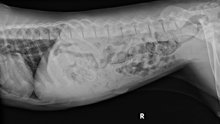
Case C: "Bella," a 5-year old mixed breed of unknown sex
.png "case-c-bella1-(1).png")
.png "case-c-bella2-(1).png")
History
Compare to X-rays taken yesterday. Patient fasted overnight. Also is still very lethargic.
Radiologist Findings
The stomach contains a moderate amount of fluid, gas, and a fusiform 6.2 x 0.9 cm structure that has a thin peripheral soft tissue rim and lucent center. This was not present/visible on the previous study. A few loops of minimally filled small bowel are present, as well as several that are moderate to severely dilated with fluid and gas. This was not present previously. There is moderate volume semi-formed feces in the colon.
Conclusions
Mango pit-type foreign object in the stomach. These are commonly not visible unless perfectly aligned with the X-ray beam. This is of a size that is too large to pass successfully. Progressive small intestinal distention, with several loops that are moderate to severely dilated consistent with segmental ileus secondary to intestinal obstruction.
Recommendations
Surgical intervention is recommended for gastric and a small intestinal obstruction.
So, how did your interpretations compare?
As much as we’d love for them to stick to treats and kibble, sometimes our pets just can’t help themselves and they end up with things like mango pits, chicken bones, and pieces of plastic in their bodies. And in those instances, we’re grateful for the powerful combination that is board certified radiologists + modern AI technology that serves as a second opinion, giving veterinary professionals the backup they need to confirm diagnoses and treatment plans.
As we anxiously await the October issue of Veterinary Practice News, where this year’s “They Ate What?!” contest winner and honorable mentions will be unveiled, we invite you to visit this page of our website to learn more about Patterson Teleradiology, powered by Vetology AI. You’ll receive evaluations in under five minutes, with the option to submit cases to a live radiologist.
You can also visit PattersonVet.com/AllThingsRad to find details about a limited time offer where you can get Vetology AI free for one month by submitting a single case through Patterson Teleradiology!
Share
Related blogs

6 Uses for AI in Vet Practices That Are Almost TOO Intelligent